DL-AAA Retinal Leakage
Retinal vascular diseases such as wet age-related macular degeneration (wet AMD), diabetic macular edema, and retinal vein occlusion are driven in large part by vascular endothelial growth factor (VEGF). A key challenge in developing new anti-VEGF therapies is evaluating their duration of action in a model with chronic, stable vascular pathology.
Model Overview
DL-alpha-aminoadipic acid (DL-AAA) is a selective Müller cell toxin. When injected intravitreally in rabbits, it induces retinal neovascularization and chronic vascular leakage that stabilizes within 10–12 weeks and persists long-term. The leakage in this model is VEGF-dependent and is reversible with anti-VEGF treatment, returning to baseline after drug clearance. This makes it particularly well-suited for comparing the efficacy and duration of action of novel anti-angiogenic and sustained-release formulations.
Typical Endpoints
- Fluorescein angiography (FA): qualitative grading and quantification of retinal vascular leakage
- Fluorophotometry: quantification of fluorescein concentration in the vitreous
- Optical coherence tomography (OCT): structural assessment of the retina
- Color fundus photography: documentation of neovascular lesion morphology
- Histology and immunohistochemistry at study termination
Representative Data
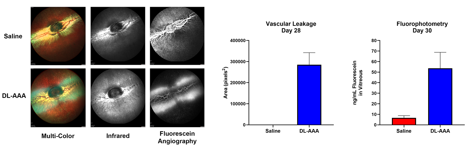
Left Column: Multi-color, infrared, and fluorescein angiography images of saline control (top) and DL-AAA treated (bottom) rabbit eyes. DL-AAA-treated eyes display pronounced retinal vascular pathology and leakage compared to controls.
Right Column: Quantification of vascular leakage area at Day 28 and vitreous fluorescein concentration (fluorophotometry) at Day 30, demonstrating significant increases in vascular leakage following DL-AAA administration.
Interested in this model?
We are happy to share more about study design options, endpoints, or timelines.
Contact us to discuss your study goals.