Retinal Vein Occlusion
Retinal vein occlusion (RVO) is a common retinal vascular disorder, second only to diabetic retinopathy in prevalence. RVO occurs when blood vessels in the retina become blocked, preventing blood flow and initiating a cascade of events that impair vision. It is a common complication of high blood pressure, high cholesterol, and diabetes. While there is no cure, current treatments to manage vision loss include intravitreal anti-VEGF injections, laser therapy, and steroid injections. Frequent dosing requirements and poor patient compliance with existing therapies drive significant demand for alternative treatment approaches.
Model Overview
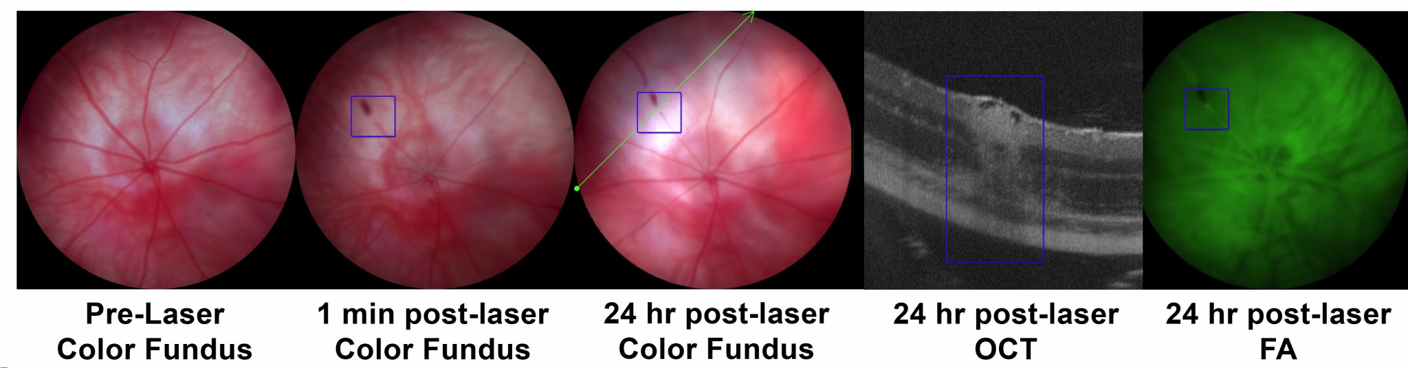
EyeCRO has validated a mouse model of RVO designed to test therapeutic intervention on retinal degeneration. Venous injection of the photo-activator dye Rose Bengal is combined with laser treatment to allow for precise and robust thrombus formation and retinal vein occlusion. Following injection of the dye, a thermal laser is used to photocoagulate up to 3 different retinal veins in a single eye.
Typical Endpoints
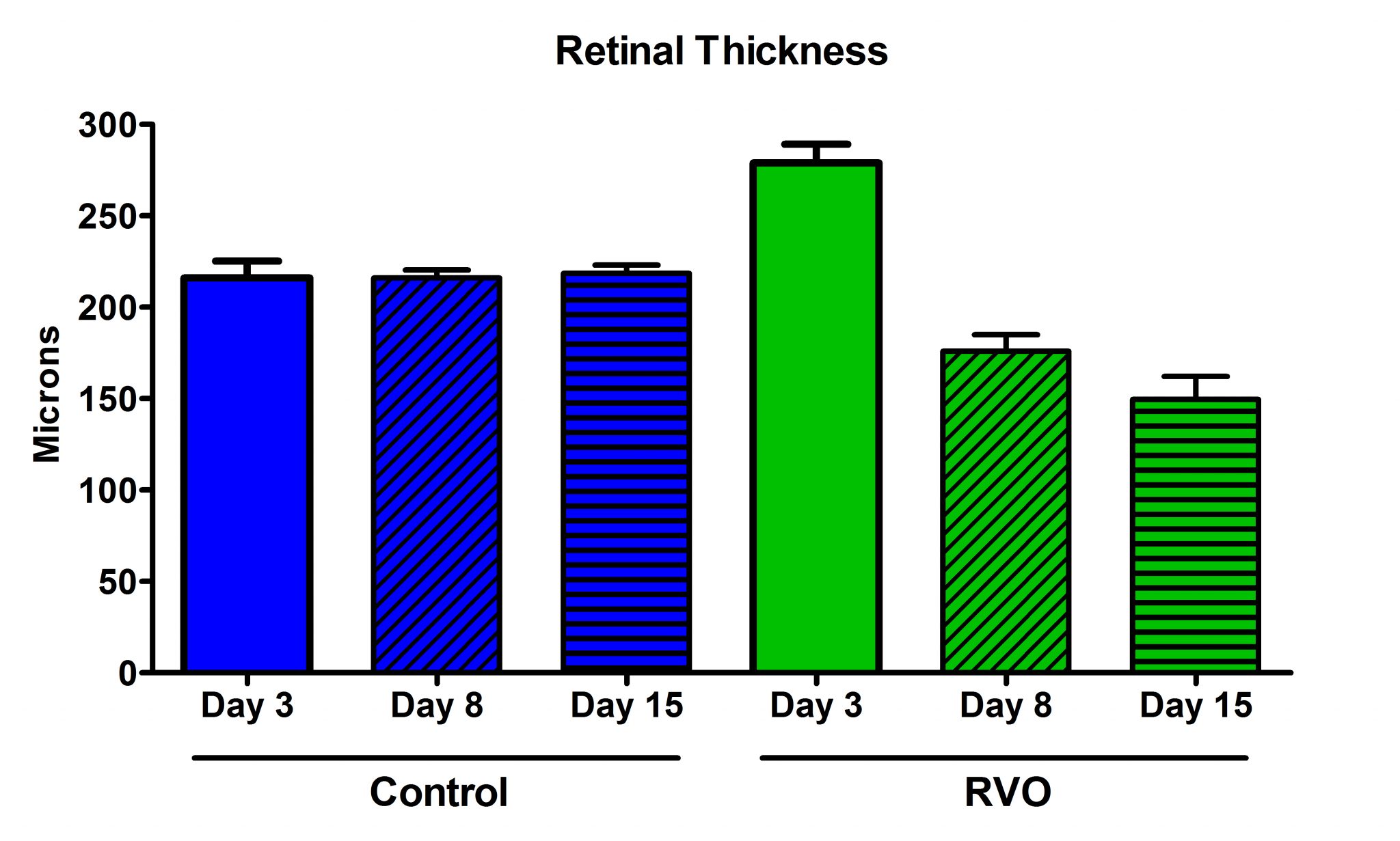
- OCT (longitudinal retinal thickness in the area of occlusion, up to 2 weeks)
- In vivo fluorescein angiography (retinal vascular leakage)
- Immunohistochemistry (retinal cell death)
- Biochemical analysis of inflammation
Representative Data
Interested in this model?
We are happy to share more about study design options, endpoints, or timelines.
Contact us to discuss your study goals.