Geographic Atrophy
Geographic atrophy (GA) is an advanced form of dry age-related macular degeneration (AMD), the most common cause of irreversible central vision loss in the elderly population. GA affects over 8 million people globally and is characterized by progressive loss of retinal pigment epithelium (RPE), photoreceptor cells, and choriocapillaris, resulting in well-defined atrophic lesions in the macula and a gradual, permanent decline in central vision.
Model Overview
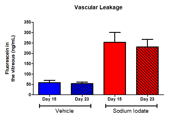
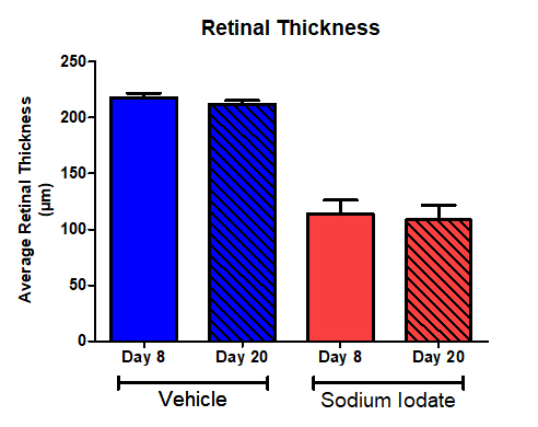
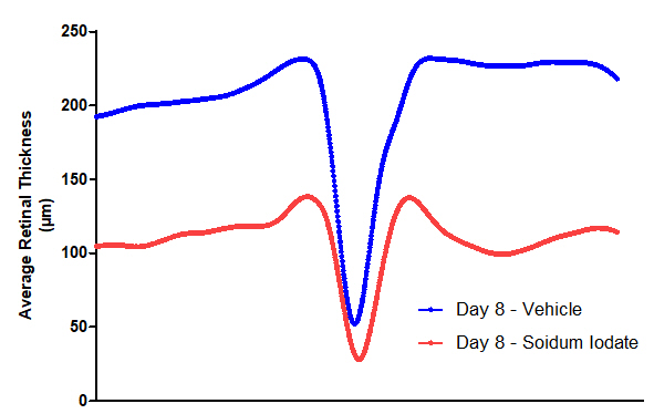
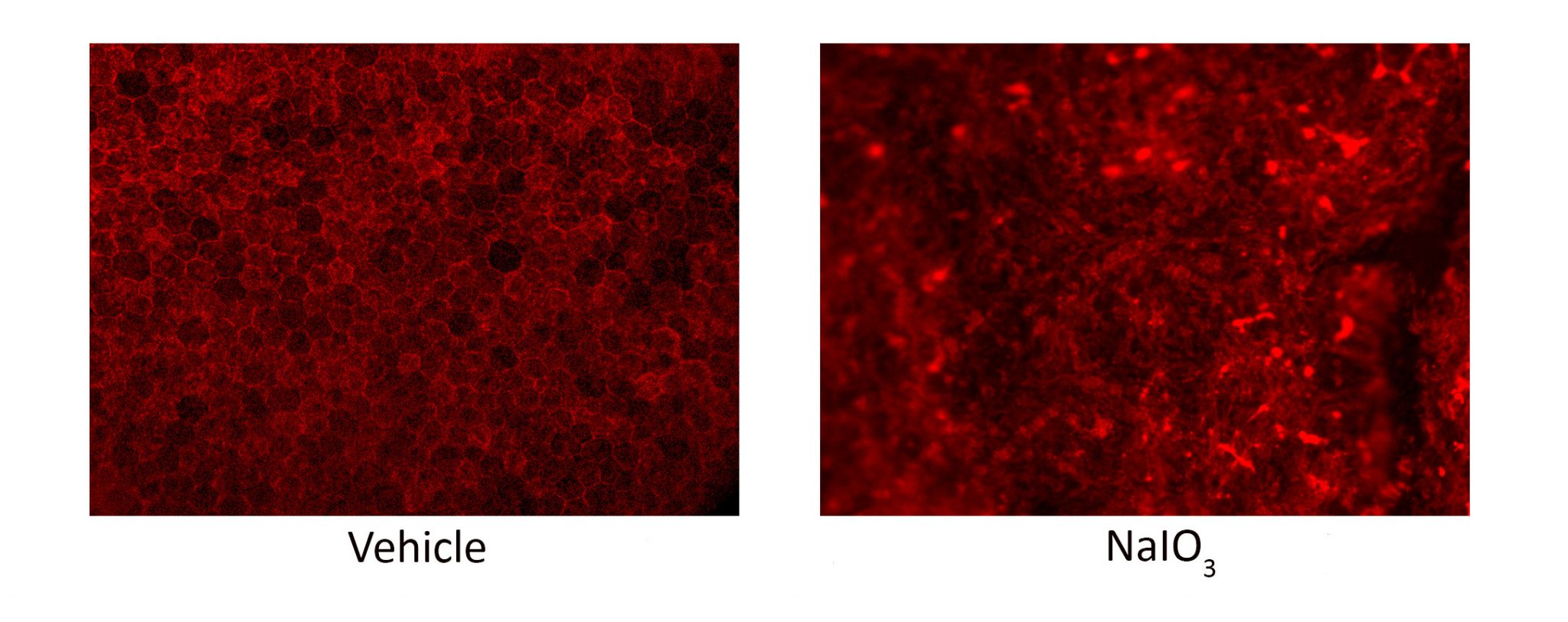
EyeCRO has established a rat model of geographic atrophy using subretinal delivery of sodium iodate (NaIO3). This toxin induces RPE necrosis through the production of reactive oxygen species, creating a focal area of RPE loss surrounded by healthy RPE that closely mimics human GA pathology. The death of RPE cells causes subsequent degeneration of photoreceptors and other retinal neurons. Atrophic lesions in this model expand over time, recapitulating the progressive nature of human GA. As the RPE maintains the outer blood-retinal barrier, RPE cell loss can be monitored non-invasively using fluorophotometry.
Typical Endpoints
- Fluorophotometry (blood-retinal barrier integrity)
- OCT imaging (retinal layer thickness, extent of degeneration)
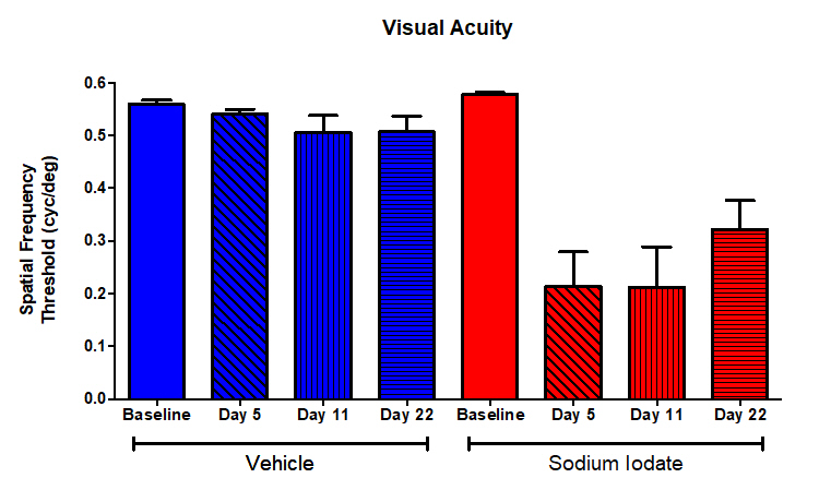
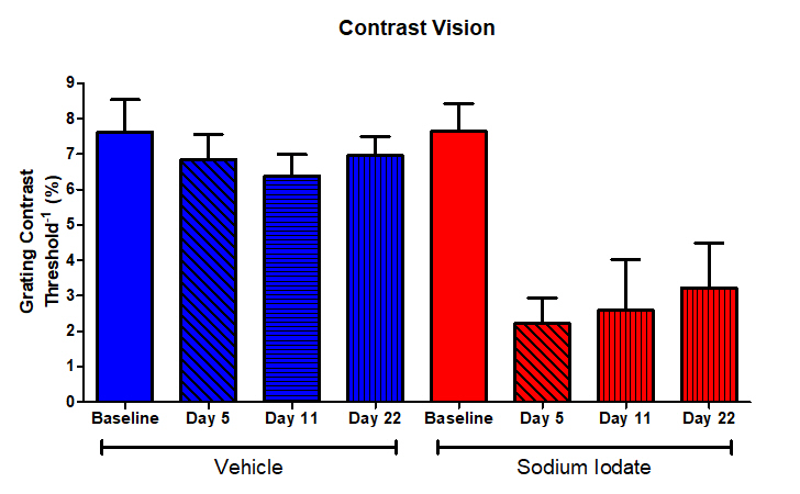
- Optokinetic tracking (visual acuity, contrast sensitivity)
- RPE flatmount analysis (RPE65 staining, atrophic area quantification)
- Histopathology and immunohistochemistry
- Microglial/macrophage infiltration (IBA1 staining)
Representative Data
Interested in this model?
We are happy to share more about study design options, endpoints, or timelines.
Contact us to discuss your study goals.