Inherited Retinal Degenerations
Inherited retinal degenerations (IRDs) are a genetically and clinically diverse group of disorders characterized by progressive dysfunction and loss of photoreceptor cells, leading to vision impairment and blindness. IRDs affect approximately 1 in 3,000 to 4,000 individuals worldwide and include conditions such as retinitis pigmentosa, Leber congenital amaurosis, and Stargardt disease. Gene therapy, neuroprotection, and cell-based therapies are actively being developed to treat these conditions, driving demand for well-characterized preclinical models.
Model Overview
EyeCRO maintains internal colonies of numerous transgenic and knockout mouse and rat lines for the study of inherited retinal degenerations (IRDs). Each line models a specific genetic mutation associated with human retinal disease.
Transgenic Models Available
| Model | Gene / Mutation | Human Disease |
|---|---|---|
| Pde6b (rd1, rd10) | Phosphodiesterase 6B | Retinitis pigmentosa |
| Rds/Prph2 | Peripherin-2 | Retinitis pigmentosa, macular dystrophy |
| RPE65 | RPE65 | Leber congenital amaurosis |
| CEP290 | Centrosomal protein 290 | Leber congenital amaurosis, Senior-Loken syndrome |
| Abca4-/- | ATP-binding cassette A4 | Stargardt disease |
| Rdh8-/- | Retinol dehydrogenase 8 | Retinal degeneration |
| Rhodopsin P23H rat | Rhodopsin | Autosomal dominant retinitis pigmentosa |
| RCS rat | Mertk | Retinitis pigmentosa |
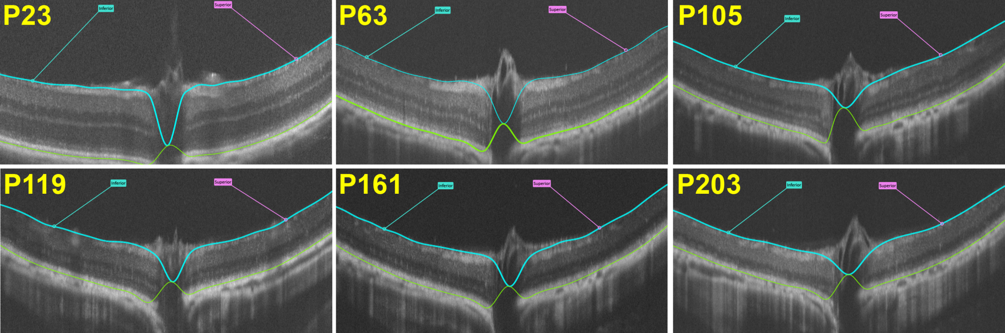
These models recapitulate key features of human Inherited Retinal Diseases (IRDs), including progressive photoreceptor degeneration and loss of visual function. Longitudinal study designs utilizing non-invasive techniques such as Electroretinography (ERG), optokinetic tracking (OKT), Optical coherence tomography (OCT), and fundus imaging allow for elegant long-term experiments to evaluate the therapeutic potential and durability of candidate treatments.
Typical Endpoints
- Electroretinography (ERG): scotopic and photopic responses
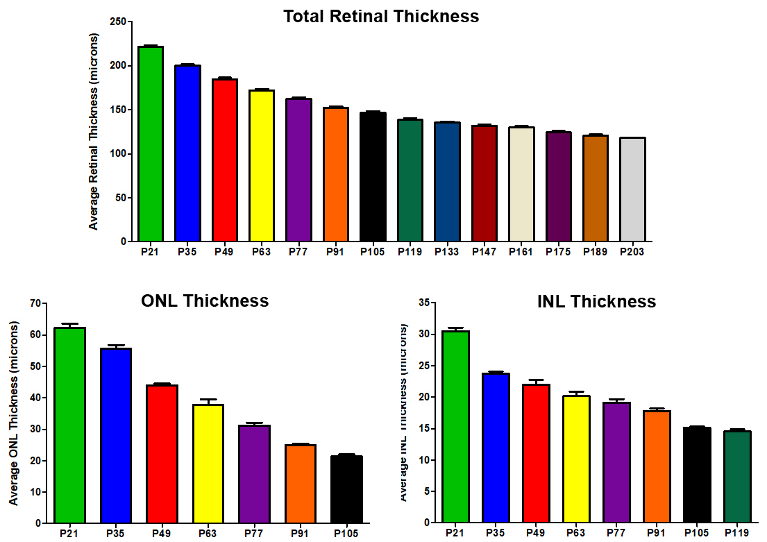
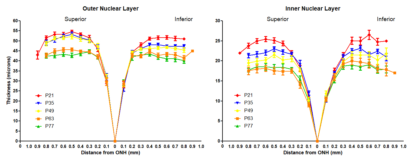
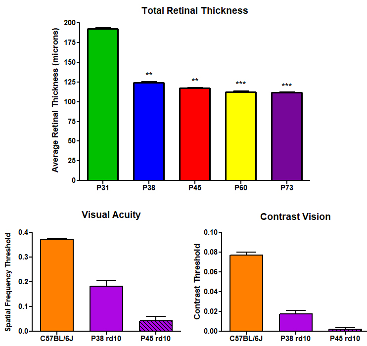
- Optical coherence tomography (OCT): retinal layer thickness over time
- Optokinetic tracking (OKT): visual acuity and contrast sensitivity
- Fundus imaging
- Histology: photoreceptor counts, outer nuclear layer thickness
- Gene and protein expression analysis
Representative Data
Interested in this model?
We are happy to share more about study design options, endpoints, or timelines.
Contact us to discuss your study goals.