Ophthalmic Imaging & Physiology
EyeCRO maintains a comprehensive suite of in vivo ophthalmic imaging and functional assessment equipment. All systems are available for mouse, rat, and rabbit studies, with trained operators and established imaging protocols across our disease model portfolio.
Our facility is equipped with both Phoenix Micron and Heidelberg SPECTRALIS imaging systems. The Micron platform enables rapid, high-quality fundus imaging, fluorescein angiography, image-guided laser application, and optical coherence tomography in small animal models. The SPECTRALIS adds advanced capabilities such as confocal SLO imaging, angiography (FA/ICG), and autofluorescence, supporting more detailed longitudinal and translational assessments.
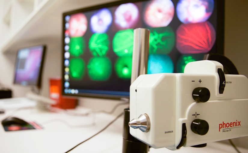
Four Micron IV devices from Phoenix Research which allow for slit-lamp imaging, color fundus imaging, fluorescein angiography, laser application, and optical coherence tomography.
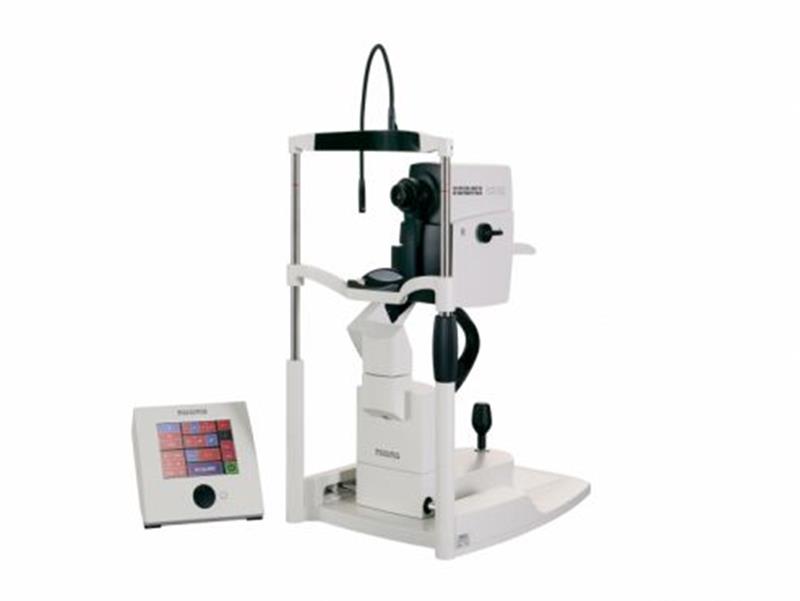
Heidelberg SPECTRALIS imaging system for confocal SLO, fluorescein/ICG angiography, and autofluorescence in preclinical ophthalmic studies.
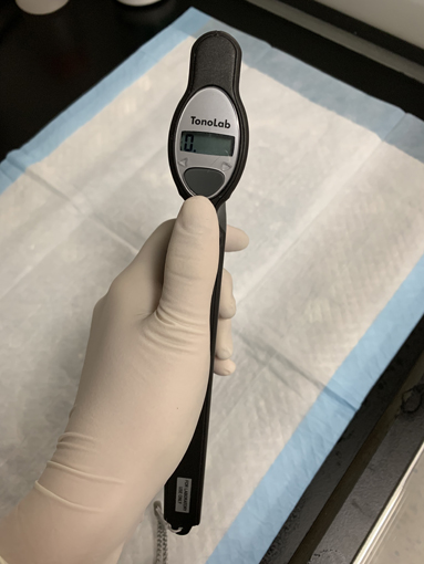
Tonolab and Tonovet devices are utilized for intraocular pressure measurments
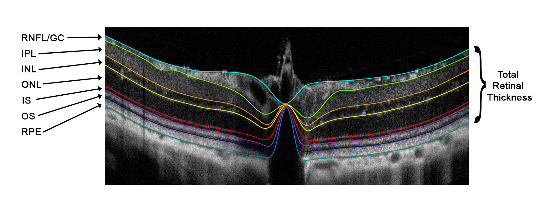
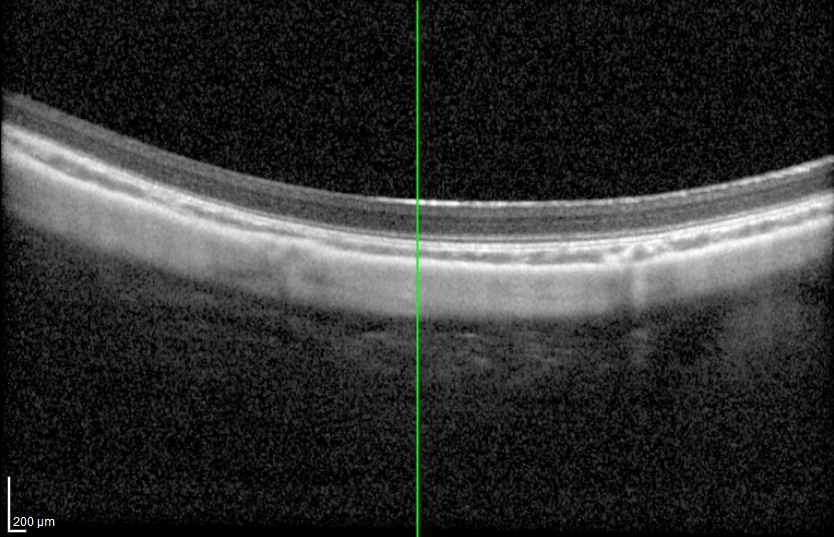
OCT Segmentations
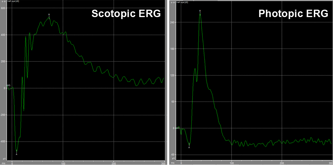
Comprehensive full-field and pattern electroretinography (ffERG, pERG) conducted on the Diagnosys Celeris system, deliver high-quality, reproducible assessment of retinal function.

Fluorophotometry studies are accomplished using the Fluorotron which allows for quantification of retinal vascular leakage and integrity of the blood-retinal barrier
A mouse pupillometer is used to measure dynamic changes in pupil diameter in response to controlled light stimuli, providing a functional readout of retinal and neurological activity.
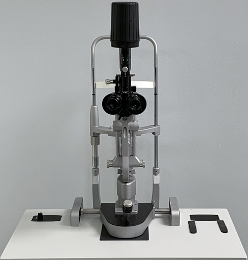
Rabbit imaging and laser application is conducted with a Haag-Streit BM900 slit lamp and ophthalmoscopic examinations are conducted with Keeler Vantage Plus and Welch-Allyn direct scope
Visual acuity and contrast vision are measured by optokinetic tracking using OptoMotry
Need specific imaging endpoints for your study?
Contact us to discuss imaging protocols, equipment availability, and endpoint selection for your next study.
Contact Us