Endotoxin Induced Uveitis
Uveitis encompasses a group of inflammatory conditions affecting the uveal tract of the eye, including the iris, ciliary body, and choroid. It accounts for 10-15% of all cases of blindness and can be associated with autoimmune diseases, bacterial or viral infections, chemical injuries, and metabolic disorders. Anterior uveitis is the most common form, accounting for the majority of the cases.
Model Overview
EyeCRO utilizes endotoxin-induced uveitis (EIU) in mice to model acute, non-autoimmune anterior uveitis. Systemic administration of lipopolysaccharide (LPS), a component of Gram-negative bacterial cell walls, triggers an innate immune-driven inflammatory response primarily in the anterior segment of the eye. Inflammation is characterized by iris hyperemia, miosis, increased aqueous humor protein concentration, and inflammatory cell infiltration into the anterior uvea and anterior chamber. Cellular infiltrate and protein levels in the aqueous humor begin to increase within 6 hours of LPS injection and typically peak at 24 hours.
Typical Endpoints
- Clinical scoring of anterior segment inflammation (iris hyperemia, miosis, fibrin, hypopyon)
- Aqueous humor cell count and protein concentration
- Inflammatory cytokine and chemokine quantification
- Histopathological evaluation of anterior uvea, ciliary body, and retina
- In vivo anterior segment imaging
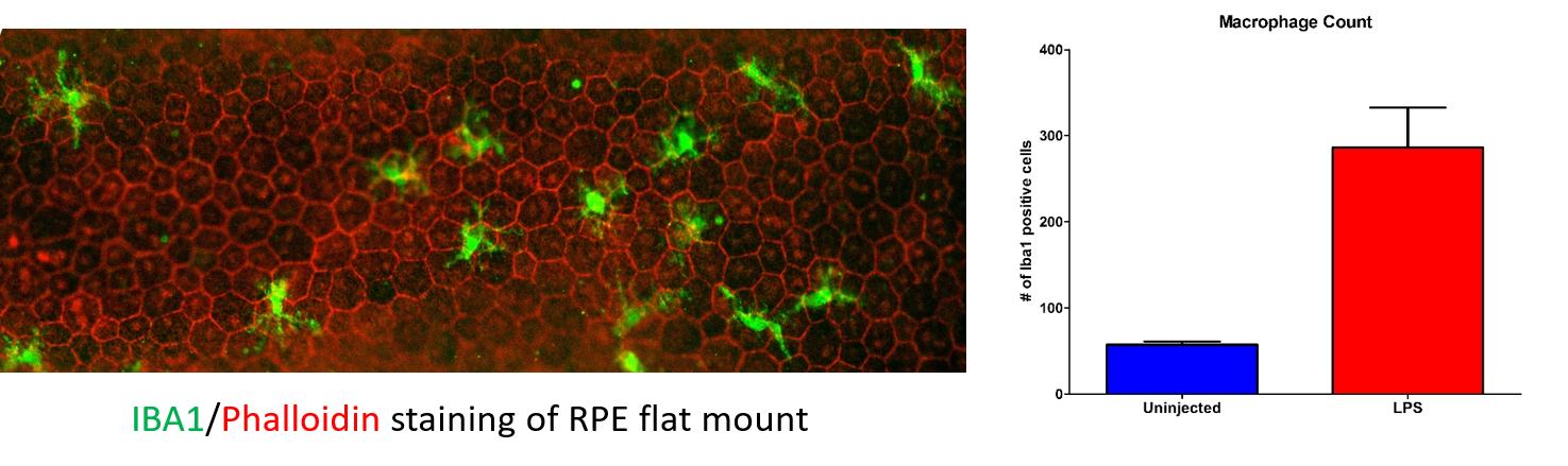
Representative Data
Interested in this model?
We are happy to share more about study design options, endpoints, or timelines.
Contact us to discuss your study goals.