Corneal Sensitivity
The cornea is the most densely innervated structure in the body. Corneal nerves mediate sensations of touch, pain, and temperature and are essential for maintaining ocular surface health through the regulation of tear production, blink reflexes, and epithelial repair.
Model Overview
Corneal nerve loss or dysfunction is frequently observed in diseases that lead to corneal opacity and vision loss. These include injury, surgical procedures, microbial infections, diabetes mellitus, and dry eye disease.
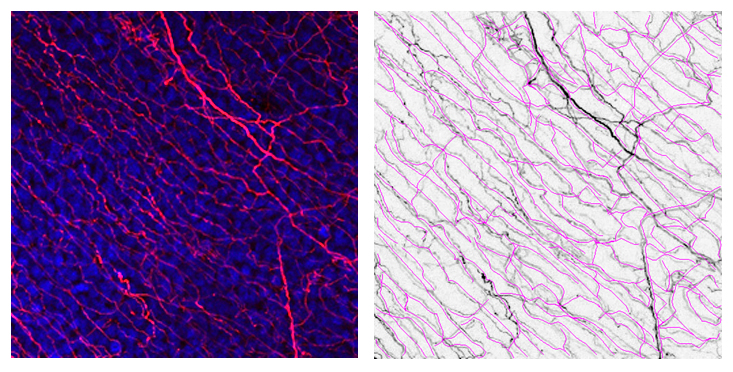
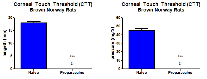
Corneal innervation can be evaluated for both function and morphology using standard instruments such as a corneal esthesiometer and β-III tubulin nerve staining.
Typical Endpoints
- Corneal mechanical sensitivity (esthesiometry)
- Nerve density and branching (β-III tubulin staining)
- Correlation with epithelial healing or surface staining
- Evaluation of nerve damage and regeneration
Representative Data
Interested in this model?
We are happy to share more about study design options, endpoints, or timelines.
Contact us to discuss your study goals.