Blue Light Damage
Intense blue light exposure induces oxidative stress and apoptosis in the retina, leading to photoreceptor degeneration. This model is utilized to investigate retinal disease mechanisms and assess neuroprotective therapies.
Model Overview
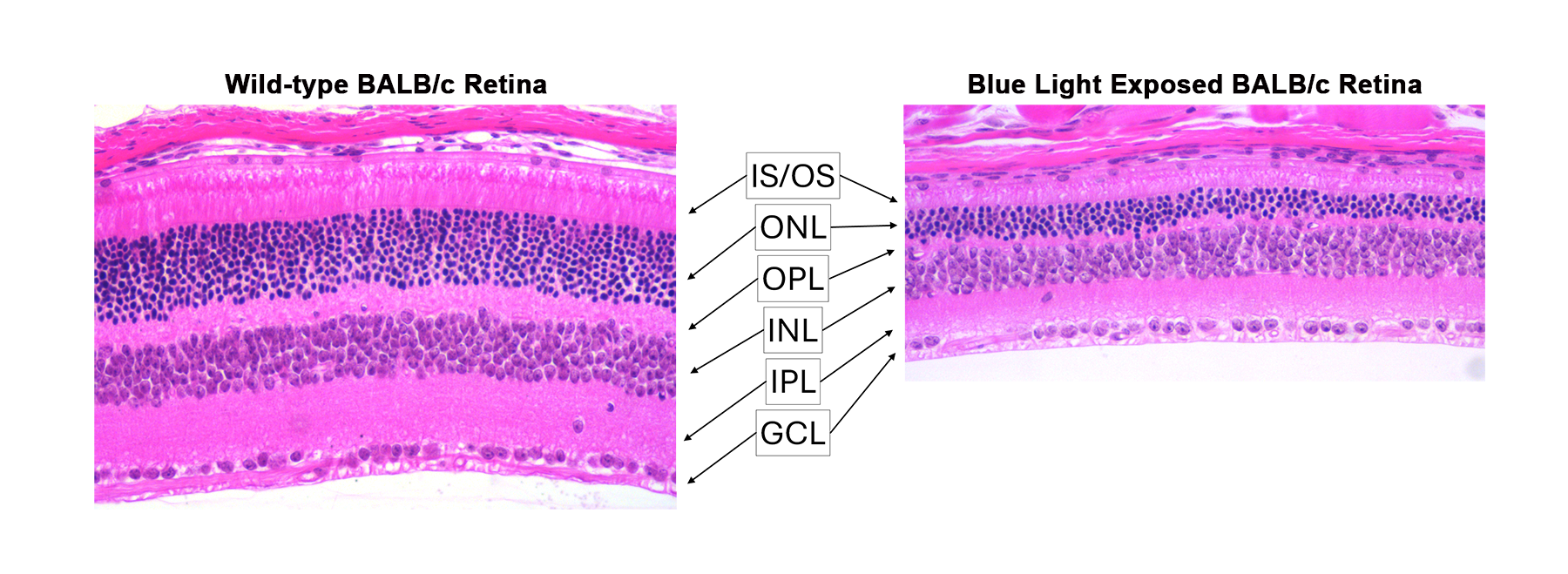
Sustained exposure to intense blue light induces rapid retinal degeneration, which can be monitored by both visual function testing and histologic analysis. This model produces phototoxic damage to the photoreceptor layer, particularly the outer nuclear layer, and is valuable for evaluating therapeutic strategies aimed at preventing retinal cell death or promoting cellular replacement.
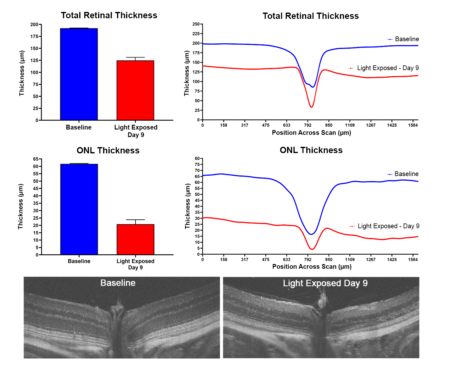
Damage can be quantified using structural measures such as retinal and ONL thickness, as well as functional assessments including electroretinography (ERG).
Typical Endpoints
- Photoreceptor layer thickness and structure (OCT or histology)
- Outer nuclear layer (ONL) thinning
- Oxidative stress markers
- Electroretinography (A-wave and B-wave amplitudes under scotopic and photopic conditions)
- Efficacy of neuroprotective or antioxidant interventions
Representative Data

Interested in this model?
We are happy to share more about study design options, endpoints, or timelines.
Contact us to discuss your study goals.